

From the age of 49 to 58, every woman should undergo mammography - X-ray of the breast - once every 3 years. This makes it possible to detect cancer at an early stage, of small size, and to start treatment earlier, which will be more effective, save life and not change its quality.
Breast cancer used to rank 1st in the world in terms of malignant diseases in women and 2nd in terms of mortality. Now earlier and more accurately detected cancer and better treated, so the prognosis for the future life of patients with breast cancer has become more favorable. And everything in this sad statistic is changing for the better every year, says Galina Yanyshevskaya, a radiologist at the Republican Clinical Medical Center.

— What is mammography and how does it differ from breast ultrasound?
— Mammography is an X-ray examination of the breast. And, like any X-ray examination, it involves radiation exposure, albeit small.
Ultrasound is an ultrasound examination. Ultrasound does not give radiation, so it can be done more often. Mammography - no, it is done according to the doctor's prescription, and the doctor also prescribes the frequency of the examination.
Both of these tests are primarily aimed at detecting malignancies in the breast. But each has its own specificity.
When a woman is young, her “mammary gland” is represented by glandular tissue. As she ages, it is replaced by fatty tissue. And the sensitivity of standard X-ray mammography at high density of breast tissue can be 48%, at low density (fat involution) - 98%, the average efficiency is 78%.
The glandular tissue is dense, and on X-ray examination some structures on the background of this dense pattern can not be seen. That is, mammography in this case is less informative than breast ultrasound. And when irradiated, a younger patient will receive a higher dose than an older patient whose mammary gland is mainly represented by fatty tissue.
So, when examining the breasts of women under 40 years of age, ultrasound is preferred. After forty, mammography is connected to ultrasound. They complement each other.
— How significant is the radiation exposure from a mammogram?
— The dose is small. But the doctor always wants to ensure that even this small risk is justified by the benefits of the trial.
When they talk about risk, I usually mention the work of our European colleagues, which shows that the risk of developing cancer is 5-6 cases per 1 million with a latency period of 10-20 years. At the same time, thanks to these studies, 330 cases of cancer are detected in the same million women. The benefit to risk ratio is about 60 to 1.
If age-related changes are already taking place in the breast, i.e. the glandular tissue is being replaced by fatty tissue, the risk is even lower. If the breast still consists mainly of glandular tissue, on the contrary, the risk is greater. For this reason, young women should not undergo mammography without a strong reason.

— At what age and how often should I get a mammogram?
— Mammograms are mandatory every 3 years for women age 49 and older. It is a preventive program to look for breast cancer. On individual recommendation and referral from a doctor, mammography can be done from the age of 40 and earlier.
Of course, not everyone wants to go for preventive examinations when their health does not suffer. But it happens that a woman has nothing to worry about, and a mammogram reveals cancer. Therefore, I urge my patients to take screening examinations responsibly.
— Do women in their 20s, 30s get mammograms?
— Cancer is getting younger, so the test is increasingly performed on younger patients. If the doctor has ordered a mammogram, it should be done, there are reasons for this. One of the reasons is that only mammography shows the presence of malignant microcalcinates. The fact is that breast cancer may not go beyond the ducts, but be widespread. Ultrasound won't show that.
When a woman chooses an active social position, is not limited to giving birth, breastfeeding children, the glandular tissue does not “work out” its tasks to the fullest extent. As a result, the involution of the mammary gland may occur not smoothly, as conceived, but by leaps, be uneven. Breast cancer begins to grow in the milk ducts and/or lobules of glandular tissue.
A malignant formation, sarcoma, can also arise from connective tissue, but it is less common.

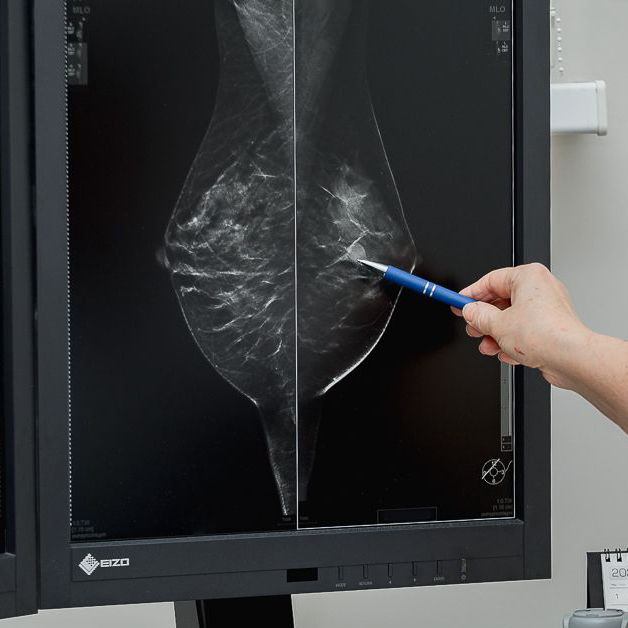
— How does mammography determine the nature of the mass: malignant or benign?
— A malignant focal (nodular) mass has irregular and indistinct contours. It deforms the surrounding pattern. If the formation has smooth and clear contours and has been stable for a long time, it is most likely benign.
If we see a focal mass but cannot determine its nature, additional tests are used: for example, contrast mammography is when a special substance called contrast is injected intravenously into the patient before the examination. If the mass is “evil”, it accumulates contrast.

Also in a situation where the doctor has doubts, tomosynthesis can be performed, which is a study that shows the breast layer by layer. The fact is that with dense tissue, the probability of seeing something is no more than 48%. And when we see the structure of the breast in more detail, the chance increases to 90%.
As you have already realized, mammography is different: standard (4 images in two projections), tomosynthesis (layer-by-layer study of the breast, or 3D-mammography), contrast mammography (contrast is introduced for additional “illumination” of malignant foci or sites).
In addition, we should not forget about ultrasound, which is also very important in case of suspected malignancy. If on mammography we see a general picture - two projections of the breast, on ultrasound we can see small details that are not visible on mammography because of the dense pattern.
A puncture may also be performed to clarify the diagnosis. As a result, we get the cells of the mass or area, and we find out whether they are “good”, “bad” or have transient, borderline characteristics.

— What should you do if you are diagnosed with cancer?
— The patient is referred to a mammologist-oncologist, a puncture is carried out, studies on organs and systems to choose the volume and tactics of effective treatment.
It is important not to panic and realize that cancer can and should be treated and you can go on living. And early treatment of cancer will allow you to maintain the quality and habitual way of life.
— What about a benign mass?
— A small, stable benign tumor can be simply observed. If it starts to grow for some reason or if the woman is concerned about the presence of the mass, it can be removed.
Small cysts can appear and “disappear” by themselves. Large cysts are punctured and the contents of the cyst are evacuated. Over time, however, the cyst may fill up again.

— How is a mammogram done? Is it painful?
— The breast is placed on the substrate and stretched out. The position is created so that it is gripped as much as possible. The breast is pressed down with the second pad. The compression necessary to take the picture is created. This takes literally a few seconds.
Then a picture is taken in two projections. The point is that a focal mass can be diagnosed only when we see it in two projections. If in one projection the focus is visible and in the other it is not, it means that it is an addition of “shadows”, a summation effect.
The procedure can be painful if the breast is represented by glandular tissue and the day of the menstrual cycle is incorrect. In menstruating women, the breast is examined on days 7-12 of the cycle, when it is relatively “calm”.
In addition, the laboratory technician applies compression gradually. If the patient is in pain, the examination is stopped. If it is tolerable, no extreme compression is created anyway, but only the compression that will make it possible to take a good picture.
On some machines, the woman herself can gradually increase the compression to the required compression - the screen indicates when enough is enough.

— How are mammograms done for women with small breasts?
— The procedure is performed in the same way regardless of whether the breasts are large or small. Mammography is also performed on men, if necessary.
— Is preparation for the study required?
— As I said before, menstruating women need to choose the right day of their menstrual cycle. Days 7 through 12 are suitable. A non-menstruating woman can do the test on any day that is convenient for her.
In addition, we recommend that you do not use deodorants with aluminum before the mammogram: they can create additional patterns (artifacts) on the image. No further preparation is required.

Talked to: Kristina Goloviychuk
Photo: Anna Zankovic
103.by