With the aim of preventive examination and early detection of mammal gland pathologies, our Center performs X-ray and morphological breast diagnostics.
INDICATIONS FOR DIAGNOSTIC EXAMINATION OF THE MAMMAL GLAND
- painful sensation;
- inflammatory changes;
- occurrence of pathologic space-occupying lesions;
- traumatic injuries;
- assessment of the condition of breast prosthesis.
DIAGNOSTICS OF SENOLOGIC DISORDERS AT THE RCMC
Mammal gland diagnostics is performed with the use of expert-class stations and, when needed, involves ultrasound-assisted needle biopsy (including vacuum-assisted biopsy performed at the EnCor ENSPIRE system):
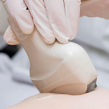
- Mammal gland ultrasonography. The structure and acoustic solidity of breasts are evaluated and, in cases of any pathologies, they are analyzed.
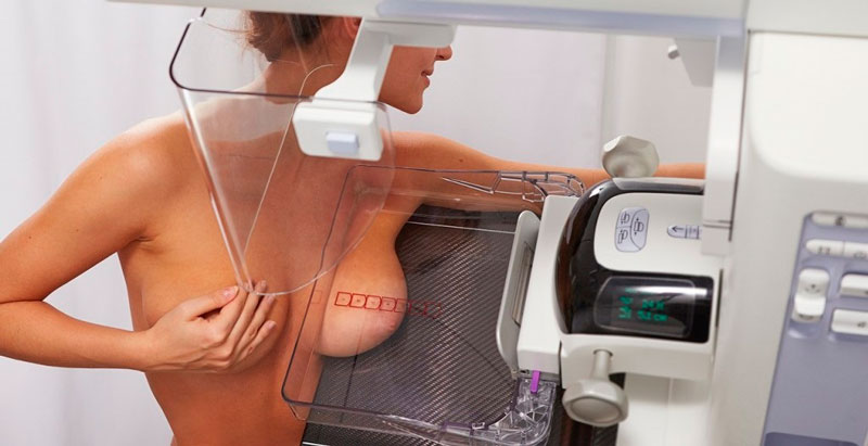
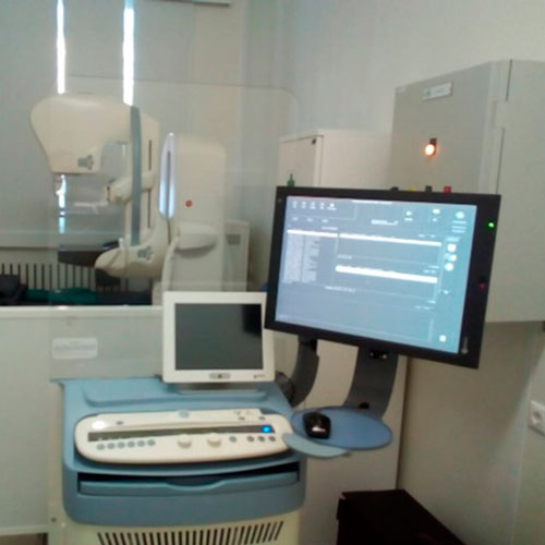
- Mammography This study is performed using a large-format digital X-ray mammography station, the most recent development in the field of mammal gland examination. The station takes full advantage of digital technologies and, together with the high imaging resolution of the X-ray system, this enables the detection of the smallest pathologies of mammal gland tissues with low exposure.

Advanced features of the station further extend its diagnostic potential:
- Contrast enhanced spectral mammography (CESM) uses X-rays of various power levels to generate two different sections. Following the introduction of the contrast medium, angiogenesis areas (i.e., areas where fine blood vessel growth occurs) are highlighted in the resulting image. This processes may potentially be related to the formation of malignant neoplasms. In this manner, CESM reduces the ambiguity of diagnosis in complicated cases, and enables physicians to diagnose breast cancer with higher certainty, accuracy and promptness to avoid costly and invasive biopsy studies.
- Tomosynthesis is a special type of mammography technique which produces a three-dimensional image of the mammal gland. The technique uses low-dose X-ray radiation emitted at varying angles. During a tomosynthesis study, the breast is placed within the station and compressed, just as during a regular mammography procedure, however the X-ray tube of the station moves around the mammal gland in an arc. The process takes only 10 seconds. The information is immediately fed to the computer which generates a 3D image of the mammal gland. The X-ray exposure during tomosynthesis is the same as during a conventional mammography.
CONTRAINDICATIONS
- Mammal gland ultrasonography: no absolute contraindications.
- Mammography: pregnancy.
PREPARING FOR THE TEST
- Mammal gland ultrasonography: no preparation is required.
- Mammography: preferably to perform on day 6-9 of the menstruation cycle.
HOW DO I HAVE THE DIAGNOSTICS AT THE RCMC?
The Center does not perform mammal gland ultrasonography as a stand-alone procedure. This examination is a part of a comprehensive breast study performed by the mammologist during a reception.
In order to have a mammography examination performed at the Centers, you will need to have a referral of your consulting physician for the examination.
To make a reservation for a visit to the mammologist (this includes mammal gland ultrasonography) or to have a mammography examination, you will need the following:
- Call the Contact Center to make an appointment
- Сonclude a contract for the provision of paid services at the registry office
- Pay the bill at the cash desk of the RCC or through the ERIP
- Come to the appointment on time.


